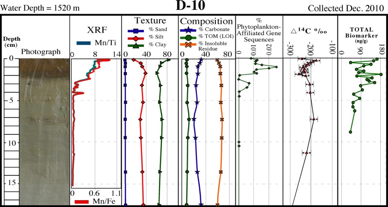
Fig 4. Core D-10 description.
Description of core D–10 collected in December 2010 showing a surficial brown layer containing dark brown-black bands corresponding to Mn spikes, and a distinct sediment texture/composition, phytoplankton-affiliated gene sequences, natural abundance radiocarbon (∆14C), and biomarkers over the surficial ~1 cm (see Fig 1 for location).

