
Figure 2. Mechanism of neutralization by human anti-CHIKV mAbs.
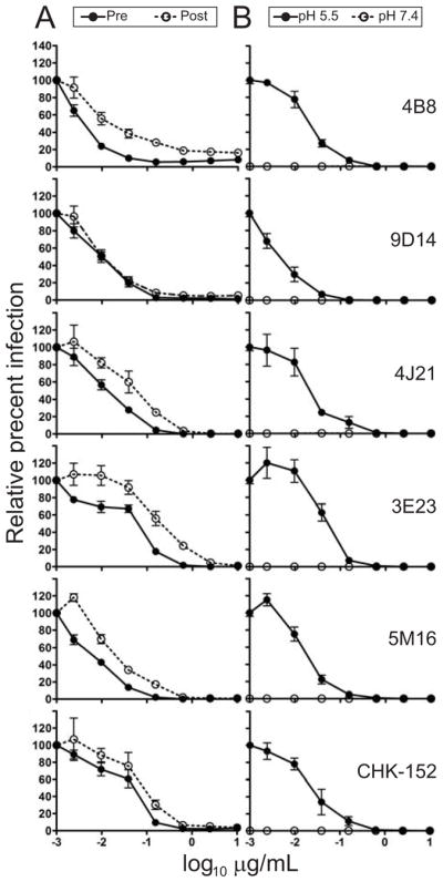
(A) Pre- and post-attachment neutralization assays. SL15649 VRPs were (i) incubated with the mAbs shown (including CHK-152, a positive control mAb) prior to addition to pre-chilled Vero cells, followed by removal of unbound virus by three washes (pre-attachment; filled circle) or (ii) allowed to adsorb to pre-chilled Vero cells followed by addition of the indicated mAbs (post- attachment; open circles). (B) FFWO assay. SL15649 VRPs were adsorbed to pre-chilled Vero cells, followed by addition of the mAbs shown (including CHK-152, a positive control murine mAb). Unbound virus was removed, and cells were exposed to low (pH 5.5 to trigger viral fusion at the plasma membrane; filled circles) or neutral (pH 7.4 as a control; open circles) pH medium at 37°C for 2 min.. For both (A) and (B) cells were incubated at 37°C until 18 h after infection, and GFP-positive cells were quantified using fluorescence microscopy. The data are combined from two independent experiments, each performed in triplicate, and represented as mean +/− SEM.