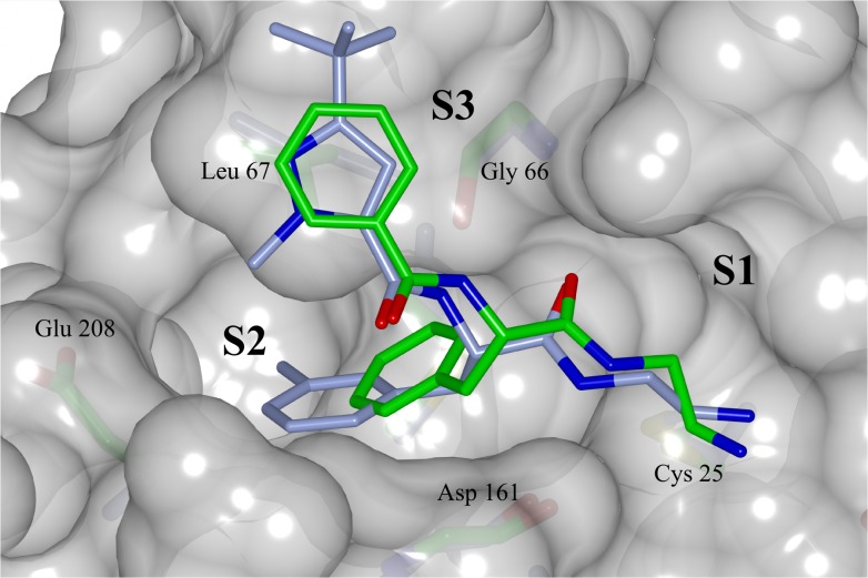
Fig 6. Superposed binding modes of compound 5 and a structural analog bound to cathepsin L (PDB code 3HHA) onto a molecular surface representation of cruzain taken from the crystal structure of 5.
Carbon atoms colored green for compound 5 in complex with cruzain and ice blue for cathepsin L analog complex. Oxygen, nitrogen, sulfur colored red, blue and yellow respectively. The binding subsites and the main residues involved at the binding are labeled. Figure prepared with CCP4mg software [52].

