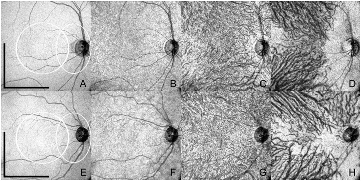
Fig 2. Enface swept-source optical coherence tomography (SS-OCT) of normal eyes of two different subjects belonging to the younger and older age groups.
The upper row (A-D) shows enface SS-OCT of a 26 year old female where the total choroidal thickness measured 346μm. Scale bars (representing A-D) = 3mm. White circular area (representing A-D): macular area. White oval area (A-D): peripapillary area. The lower row (E-H) shows enface SS-OCT of a 47 year old female where the total choroidal thickness measured 201μm. Scale bars (representing E-H) = 3mm. White circular area (representing E-H): macular area. White oval area (E-H): peripapillary area. Retinal pigment epithelium (A and E) and individual choroidal layers, namely choriocapillaris (B and F), inner choroid (C and G) and outer choroid (D and H) have distinct features. Note variable choroidal thicknesses throughout the posterior pole, revealed by the visualization of the sclera in some areas, more commonly in the peripapillary region and temporal to the macula.

