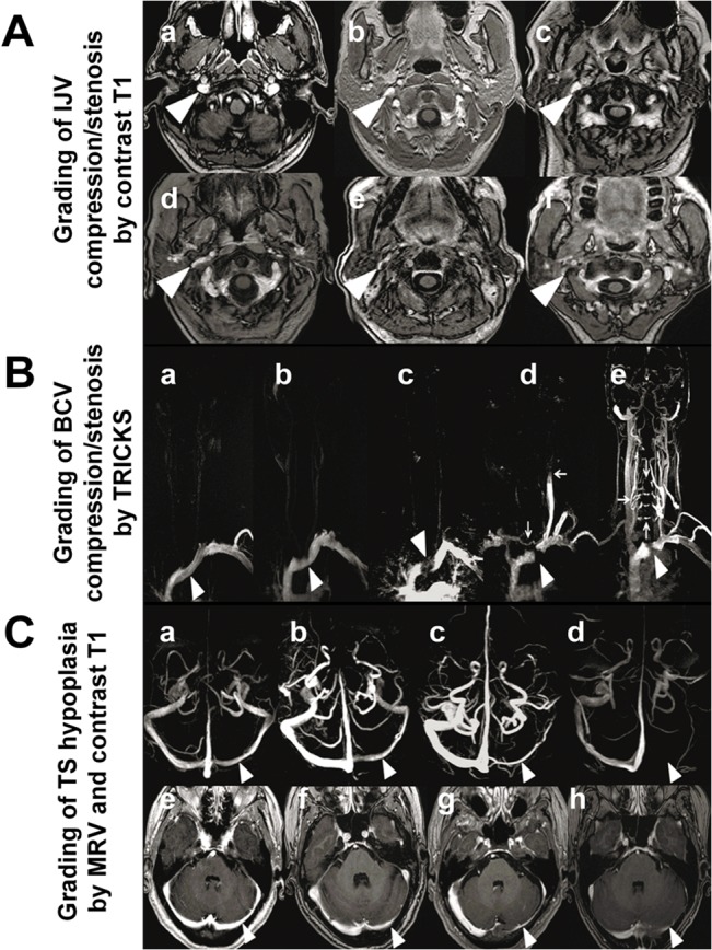
Fig 1. Grading of compression/stenosis using Contrast-enhanced MR (magnetic resonance) imaging.

(A): Grading of IJV (internal jugular vein) compression/stenosis by Contrast T1 (contrast-enhanced axial T1-weighted MR imaging). Grade 0: normal round or ovoid (a), Grade 1: mild flattening (b, c); Grade 2: moderate flattening (d); Grade 3: severe flattening (e), pinpoint (f) or not visualized. (B): Grading of left BCV (brachiocephalic vein) compression/stenosis by TRICKS (time-resolved imaging of contrast kinetics). Grade 0: normal (a, arrowhead), Grade 1: BCV with mild filling defect by the aortic compression (b, arrowhead); Grade 2: left BCV interrupted at the aortic arch (c, arrowhead) with filling defect, but without collateral; Grade 3: left BCV compression/occlusion (d-e, arrowhead) with different types of venous collaterals filling and reflux: Venous flow drains across the midline into the right IJV through the anterior cervical veins from left subclavian vein (d, vertical arrow); Reflux of IJV (d, horizontal arrow), the contrast medium injected from left subclavian vein appeared retrograde into left IJV; Collaterals of vertebral venous system, presence of collaterals of vertebral venous system, from the left subclavian vein draining directly through intrarachidian anastomoses to contralateral side at different levels (e, arrows). (C): Grading of TS (transverse sinus) asymmetry by MRV (magnetic resonance venography) (C a–C d). Grade 0: symmetrical TS (a); Grade 1: TS asymmetry≤50%(b); Grade 2: TS asymmetry >50% (c); and Grade 3 aplasia or signal absent (arrowhead pointing locations for comparison) (d). Grading of TS asymmetry by Contrast T1(C e–C h). Grade 0: symmetrical TS (e), Grade 1: TS asymmetry≤50% (f); Grade 2: TS asymmetry>50%(g); and Grade 3:aplasia or signal absent(arrowhead pointing locations for comparison) (h).
