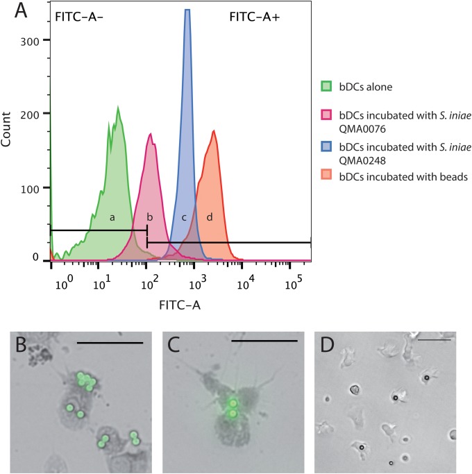
Fig 6. Phagocytic capability of bDCs.
(A) (a) Histograms showing barramundi dendritic cells alone, in presence of stained bacteria (b) QMA0076 and (c) QMA0248 and (d) in presence of fluorescent beads. Note that ingestion of beads and bacterial cells shifts fluorescence of DCs fluorescence significantly the right along the x-axis (z > 1.96) in b, c and d when compared to a. (B-C) Fluorescent microscopy and (D) inverted light microscopy of dendritic-like cells incubated with fluorescent beads. Scale bars represent 50 μm.

