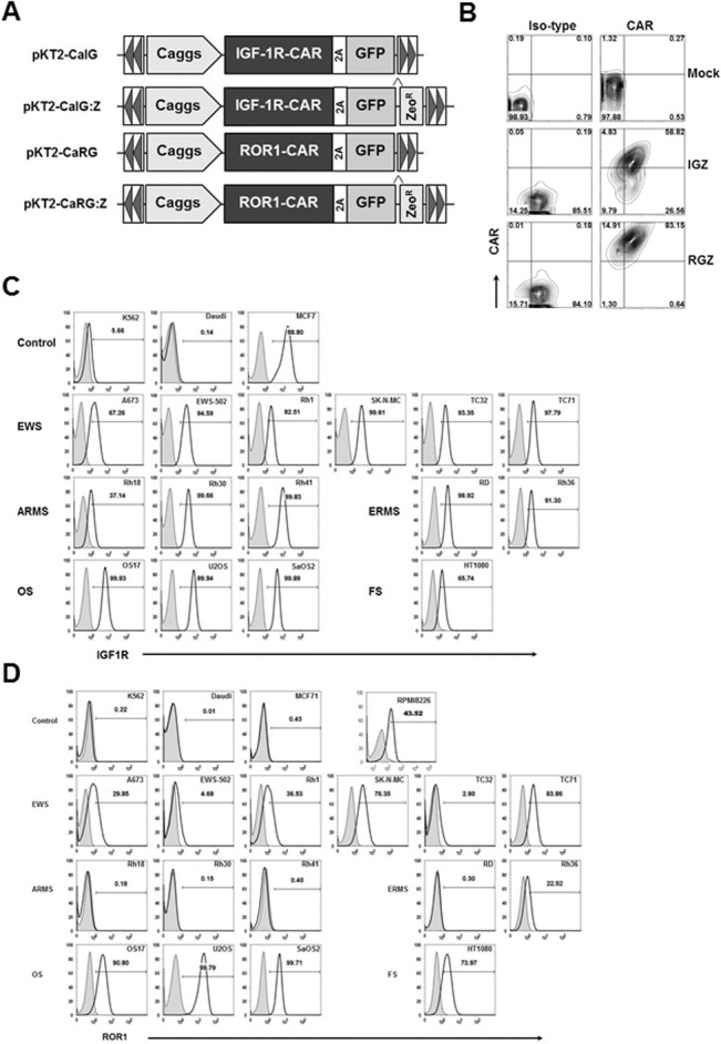
Fig 1. Cell surface expression of chimeric antigen receptor (CAR) and GFP after transfection and expression of IGF1R and ROR1 in a panel of sarcoma cell lines.

(A) Schematic representation of the Sleeping Beauty (SB) transposons encoding IGF1R CAR and GFP (pKT2-CaIG), IGF1R and GFP:Zeocin (pKT2-CaIG:Z), ROR1 CAR and GFP (pKT2-CaRG), and ROR1 CAR and GFP:Zeocin (pKT2-CaRG:Z). The CAR contains the 4-1BB signaling domain (not shown). A Caggs promoter and a “self-cleaving” 2A peptide flanking the CAR sequence were used to regulate co-expression of CARs and GFP or GFP-Zeocin fusion in the SB transposon trans vectors, namely pKT2-CaIG, pKT2-CaRG, pKT2-CaIG:Z, and pKT2-CaRG:Z. (B) Expression of CAR and GFP in T cells derived from a sarcoma patient (Patient 1) after transfection of PBMCs with pKT2-CaIG:Z/pCMV-SB100X or pKT2-CaRG:Z/pCMV-SB100X or no DNA (as mock) and selection with zeocin. Similar data were obtained in T cells from other two patients with sarcomas and two healthy donors (data not shown). (C) Flow cytometric analysis of IGF1R expression in sarcoma cell lines including Ewing sarcoma (EWS), alveolar or embryonal rhabdomyosarcoma (ARMS or ERMS), osteosarcoma (OS), and fibrosarcoma (FS). K562 (erythroleukemia), Daudi (B-cell Burkitt lymphoma), MCF7 (breast cancer) cell lines were used as control. (D) Flow cytometric analysis of ROR1 expression in sarcoma cell lines. RPMI8226 (multiple myeloma) cell line was used as control. Similar data shown in (C) and (D) were obtained in at least three independent assays.
