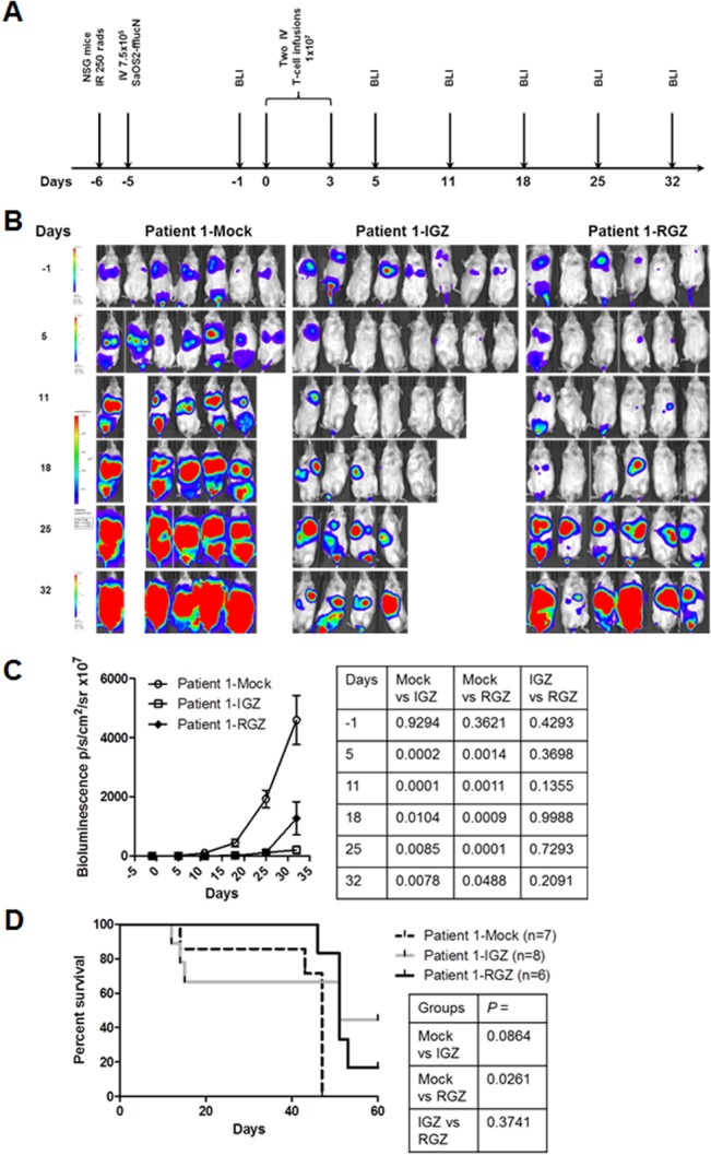
Fig 4. In vivo anti-sarcoma activity of IGF1R and ROR1 CAR T cells in a disseminated mouse model.

(A) The experimental schedule of tumor cell injection, CAR T cell infusion and BLI monitoring. Prior to testing, all mice displayed normal healthy status. B) Bioluminescent imaging (BLI) of tumor growth in NSG mice (three groups, n = 6–8 each) treated with a sarcoma patient derived T cells expressing IGF1R CAR (IGZ), ROR1 CAR (RGZ) or mock T cells. SaOS2-fflucN cells were transduced with a lentiviral vector expressing humanized firefly luciferase and truncated nerve growth factor receptor (NGFR). Two mice in the mock group died of tumor progression on day 8. Four mice in IGZ group died of unknown causes on day 8, 9, 16 and 24, probably due to cytokine storms. (C) Bioluminescent intensity of the mice treated with the T cells. All p values were shown in the right panel table and were verified independently. (D) Animal survival after T-cell therapy. All p values were determined using Mantel-Haenszel logrank test and shown in the right panel table. The p values were independently confirmed. Note that p > 0.05 between mock vs IGZ was likely due to a small sample size.
