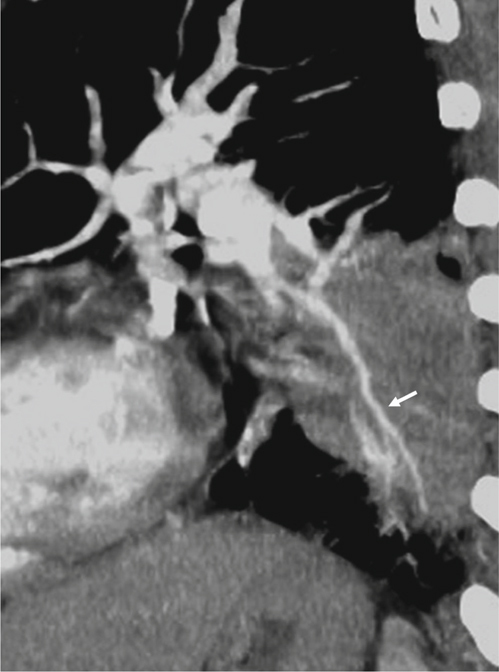
Fig. 3.

Penetration. This is a 64-year-old male with a mass lesion in the left lower lobe. He received ultrasonographyguided biopsy, and pathology revealed adenocarcinoma. Sagittal reformatted CT image reveals the pulmonary artery passes through the lesion without change of the vascular course or caliber of the pulmonary artery (white arrows).
