
Figure 2.
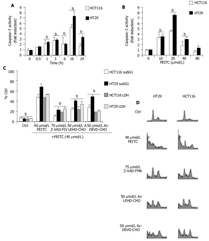
A: Time dependant Induction of caspase -3 activities in HCT116 and HT29 cells treated with 40 µmol/L PEITC as determined at 0.5, 1, 3, 6, 16 and 24 h. B: Concentration dependant activation of caspase-3 as determined at 24 h after incubation with PEITC (10-80 µmol/L). C: Z-VAD-FMK, Ac-LEHD-CHO and Ac-DEVD-CHO inhibition on PEITC mediated apoptosis as determined at 24 h. bP < 0.01, significant difference compared to control groups. Cells were treated with individual caspase inhibitors for 1 h prior to the addition of 40 µmol/L PEITC. SubG1 populations were determined by propodium iodide staining followed by flow cytometry analysis as described in the materials and methods. bP < 0.01, comparing PEITC (alone) with control groups. D: Representative flow cytometry analysis of HCT116 and HT29 cells was used to determine the extent of DNA fragmentation in cells treated with PEITC (40 µmol/L) in the presence of caspase inhibitors. Data were determined by PI staining at 24 h. Figure shows representative example of at least 3 experiments. In each analysis, 10 000 events were recorded. bP < 0.01, PEITC (alone) vs control and caspase inhibitor treated groups.