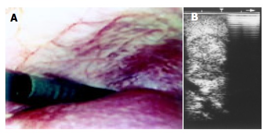
Figure 6.

A: Upper aspect of right lobe of the liver. The surface is not perfectly smooth owing to the presence of alternately reddish areas slightly elevated with respect to lighter colored ones; B: Laparoscopic sonogram showing a very well defined tumor near a vascular structure.
