Figure 8.

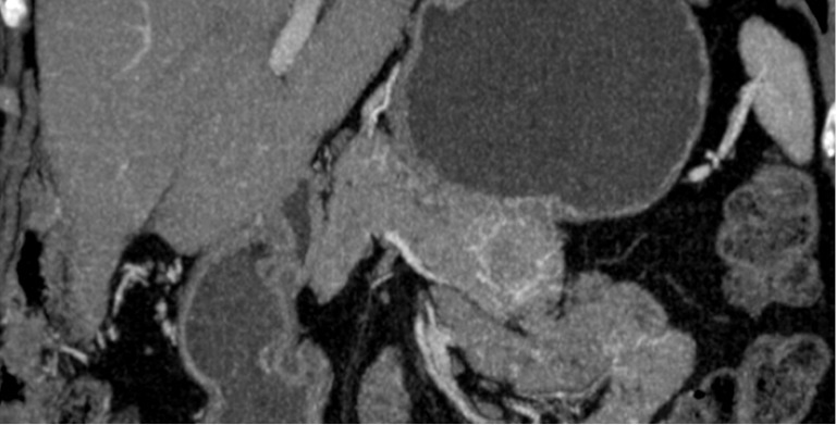
CT findings of SCN. (A) Axial image. Note the septa coming from the central scar; (B) sagittal image. Note the focal high-intensity lesion within a cyst representing hemorrhage (arrow). CT, computed tomography; SCN, serous cystic neoplasm.

CT findings of SCN. (A) Axial image. Note the septa coming from the central scar; (B) sagittal image. Note the focal high-intensity lesion within a cyst representing hemorrhage (arrow). CT, computed tomography; SCN, serous cystic neoplasm.