
Figure 3.
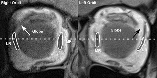
Coronal magnetic resonance imaging near the equator of the globe in an elderly patient with age-related divergence insufficiency esotropia shows an intact lateral rectus-superior rectus band in the right orbit (white arrow) with a normal lateral rectus pulley position and a dehisced lateral rectus-superior rectus band in the left orbit (black arrow) with an inferiorly displaced lateral rectus pulley. MR = Medial rectus, LR = Lateral rectus