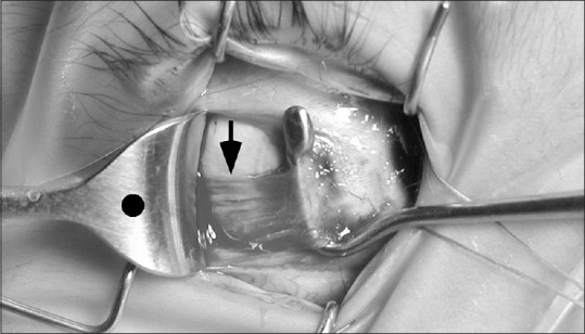
Figure 4.

Intraoperative photo shows the lateral rectus muscle insertion is in the correct temporal anatomic location, but the posterior muscle belly is inferiorly displaced by one-half tendon-width near the equator (black arrow). The posterior muscle should be centered on the retractor, marked with a black dot
