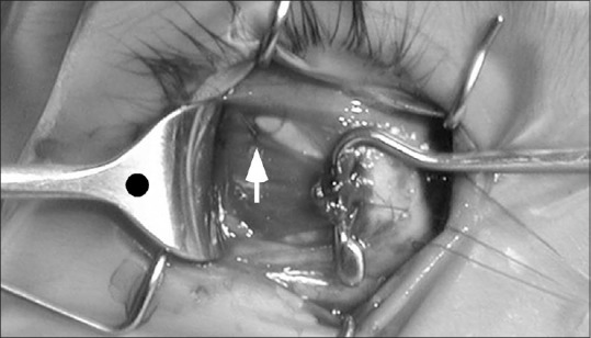
Figure 5.

Intraoperative photo of the lateral rectus equatorial myopexy procedure shows a single 6–0 polyester monofilament suture is placed through the equatorial sclera and adjacent superior lateral rectus muscle belly (white arrow) to lock the posterior muscle belly into its correct temporal location, centered on the black dot marking the center of the retractor
