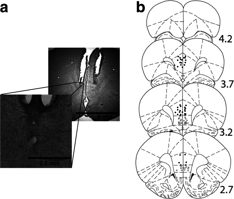
Fig. 2.
a Photograph of a representative placement which illustrates the area around the injection. There is no obvious indication of gliosis as a result of the microinfusions. b Approximate locations of infusion cannula tips, in the prelimbic (black dots) and infralimbic (grey dots) sub-regions of the mPFC. The placements on the borderline with the prelimbic area but which were not clearly infralimbic are also shown in black. Placements are shown on coronal plates adapted from Paxinos and Watson (1998), with numbers indicating distance from bregma in millimetres

