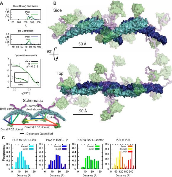
Figure 7. Interdomain arrangement in PICK1LKV tetramer.
(A) EOM on decomposed tetramer form factor of PICK1LKV. Fit of optimal ensemble to combination of PICK1LKV samples at 2.3 and 3.5 mg/ml. Top panels show Dmax and middle panel Rg of generated pools in blue and selected pools in green. Bottom panel shows fit to SAXS form factor. (B) Illustration of flexibility of PICK1LKV. The BAR domain is shown in cyan/blue. PDZ domain is green and the transparency reflects frequency of conformation in optimal ensemble. Linker region is in purple. The C-terminal is removed for visual clarity. (C) Histograms of sampled distances between the PDZ domain and the position of the linker on the BAR domain (light blues), the tip of the BAR domain (dark blues), and the center of the BAR domain (greens) and between the PDZ domains within the dimer (orange), as well as between the two distal PDZ domains (red) and the central PDZ domains (yellow) (100 ensembles, bin size 20 Å).

