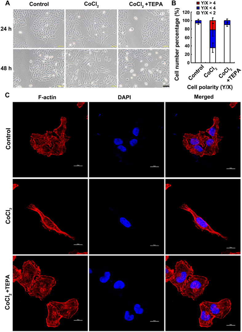
Figure 3. TEPA inhibits the CoCl2-induced morphologic changes of MCF-7 cells.
(A) Few spindle-shaped cells were observed in cells treated with 50 μM TEPA supplement. Scale bars = 100 μm. (B) The morphology of MCF-7 cells was evaluated using polarity assay by comparing the ratio of major axis/minor axis. (C) Cell morphology was further evaluated by cell cytoskeletal staining. Cells were stained with TRITC-phalloidin which is specific for cellular F-actin. Scale bar= 50 μm.

