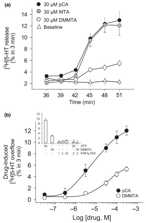
Fig. 7.
Releasing effect of DMMTA from rat brain hippocampal slices preloaded with [3H]5-HT. Slices were prepared, labeled with the radioactive tracer and exposed to drugs in superfusion. DMMTA, MTA or pCA were added at t = 39 min of superfusion and maintained until the end of the experiments. 6-Nitro-quipazine (6-NO2-QUI) was introduced at t = 30 min. Panel (a) Time course of the release of [3H]5-HT from slices exposed to 30 μM DMMTA, MTA or pCA. Data are expressed as FRR (see Materials and methods). Panel (b) and inset. Concentration–response curve of the effects of DMMTA or pCA and block by 6-NO2-QUI. Data are expressed as drug-induced [3H]5-HT overflow, calculated by subtracting the basal release (second fraction collected; t = 36–39 min) from the release measured in the fifth fraction collected (t = 45–48 min), where the drugs generally reached maximum effect. The data presented are mean ± SEM of 4–6 experiments in triplicate. *p < 0.001 compared to the effect of pCA or DMMTA alone (two-tailed Student’s t-test).

