
Fig. 3.
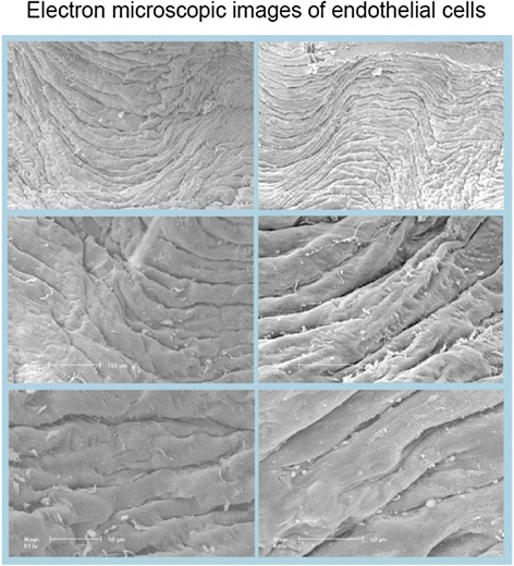
Representative scanning electron micrographs of the endothelium of human internal arteries after exposure to solvent (left) and after exposure to 10 μM rapamycin (right) for 20 h taken from the same patient. Different magnifications are indicated by bars of 200, 100 and 50 μm (from top to bottom). Some depositions of blood cells and noncellular material are seen in both groups. However, the endothelial layer is well preserved in both groups, i.e. there were no differences between rapamycin-exposed and control rings