Fig. 4.

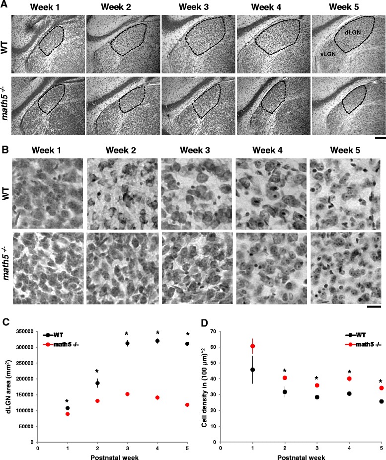
Cytoarchitecture of dLGN in WT and math5−/−. a Coronal sections of dLGN stained for Nissl at different postnatal weeks in WT (top panels) and math5 −/− (bottom panels). In math5 −/−, dLGN boundaries are well delineated, but the nucleus is smaller compared to WT (vLGN: ventral lateral geniculate nucleus). Scale bar = 200 μm. b High power images of Nissl stained dLGN cells at different postnatal weeks in WT (top panels) and math5 −/− (bottom panels). Scale bar = 20 μm. c Scatter plot depicting the mean dLGN area ± SEM as a function of postnatal week in WT (black) and math5 −/− (red). Compared to WT, the dLGN of math5 −/− was smaller at all weeks (*, weeks 1, 3–5 p <0.0001; week2 p <0.01). d Scatter plot showing cell density (±SEM) as a function of postnatal week in WT (black) and math5 −/− (red). Compared to WT, cell density was significantly higher in math5 −/− in weeks 2–5 (*, Student’s t-test, p <0.003)
