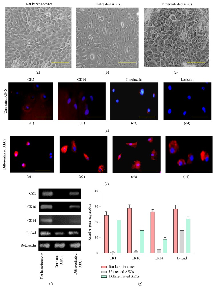
Figure 4.
Differentiation potential of amniotic membrane epithelial cells into keratinocyte-like cells. Cytomorphology of (a) positive control, (b) untreated AECs, and (c) induced keratinocytes-like cells. Immunofluorescence staining with CK5, CK10, involucrin, and loricrin in ((d1)–(d4)) untreated AECs and in ((e1)–(e4)) induced cells. Expression of mRNA of CK1, CK10, CK14, and E-Cadherin in positive control and untreated and treated cells has been shown in (f). Expression of these genes was significantly upregulated in treated group as compared to untreated AECs. (g) showed graphical representation of RT-PCR analysis.

