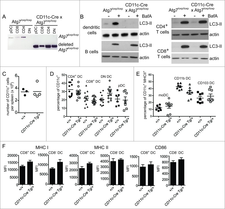
Figure 3.
Autophagy is not required for DC development or homeostasis. (A) Atg7-WT or Atg7-DC CKO CD8+ DC, CD4+ DC, CD8− CD4− double-negative (DN) DC and plasmacytoid DC (pDC) were isolated from spleen. Genomic DNA was isolated and PCR performed for the intact loxp-flanked Atg7 allele and the deleted Atg7 allele. (B) Purified Atg7-WT or Atg7-DC CKO DC, B cells, CD4+ T cells and CD8+ T cells were cultured overnight and incubated in the presence or absence of BafA for 2 h. Cell lysates were subjected to SDS-PAGE and immunoblotted for LC3 and actin. (C) CD11c+ DC were purified from Atg7-WT or Atg7-DC CKO spleens. Bar indicates mean. Data pooled from 3 independent experiments. (D) Proportions of CD11c CD4+ DC (CD4 DC), CD11c+ CD8+ DC (CD8 DC), CD11c+ CD4− CD8− DC (DN DC) and CD11c+ CD45RA+ plasmacytoid DC (pDC) present in Atg7-WT or Atg7-DC CKO spleens. Bar indicates mean, each circle represents an individual mouse. (E) Proportions of monocyte-derived DC (moDC), CD11c+ CD11b+ DC (CD11b DC) and CD11c+ CD103+ DC (CD103 DC) present in Atg7-WT or Atg7-DC CKO lung parenchyma. Gating strategies for respiratory tract DC are described in Materials and Methods. Bar indicates mean, each circle represents an individual mouse (F) Expression levels of MHC I, MHC II, and CD86 for immature CD8+ DC and immature CD8− DC isolated from Atg7-WT or Atg7-DC CKO spleens. Data are mean ± 1 SEM, n = 3 or 4 mice per group.

