Figure 2.
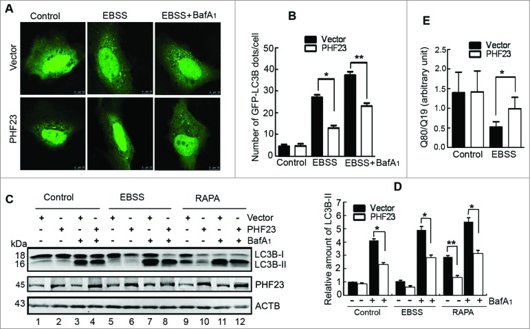
PHF23 overexpression impairs EBSS-induced autophagosome formation. (A) Representative confocal microscopy images of GFP-LC3B distribution obtained from U2OS cells cotransfected with the indicated plasmids and treated with 10 nM bafilomycin A1 (BafA1) and/or EBSS for the last 2 h. (B) Quantification of GFP-LC3B puncta per cell treated as in (A). Data are means ± SD of at least 100 cells scored (*P < 0.05, **P < 0.01). (C) U2OS cells were transfected as indicated for 24 h, treated with 10 nM of BafA1 and/or EBSS or rapamycin (RAPA, 2 μM) for the last 2 h. The levels of LC3B-II were detected by protein gel blot. (D) Quantification of LC3B-II levels relative to ACTB in cells treated as in (C). Average value in vector-transfected cells without BafA1 treatment was normalized as 1. Data are means ± SD of results from 3 experiments (*P < 0.05, **P < 0.01). (E) U2OS cells were cotransfected with polyQ80-luciferase (or control polyQ19-luciferase), vector (or PHF23) as indicated for 30 h, and treated with or without EBSS for the last 2 h. Luciferase activities were monitored, and polyQ80-luciferase/polyQ19-luciferase ratios were calculated (*P < 0.05)..

