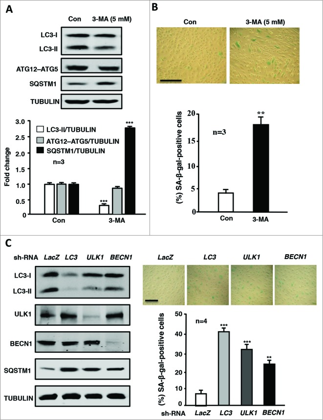
Figure 5.
Inhibition of autophagy promotes endothelial senescence. Young HUVECs were cultured in serum-growth medium in the absence (Con) or presence of the autophagy inhibitor 3-MA (5 mmol/L) for 5 d. The cells were then subjected to (A) immunoblotting analyses of LC3-I/-II, ATG12–ATG5, SQSTM1, and tubulin, which served as a loading control. (B) SA-β-gal staining. (C) The young HUVECs were transduced either with rAd/U6-LacZshRNA as control or rAd/U6-LC3shRNA or rAd/U6-ULK1shRNA or rAd/U6-BECN1shRNA. Five d post transduction, cells were subjected to immunoblotting analyses of LC3, ULK1, BECN1, SQSTM1, and tubulin (left) and senescence-associated (SA)-ß-gal staining (right). Quantification of the signals is shown in the bar graphs in the corresponding panels (n = 3 or n = 4 as indicated in the figures). **P < 0.01, *** < 0.001 vs. control (Con) or LacZshRNA.

