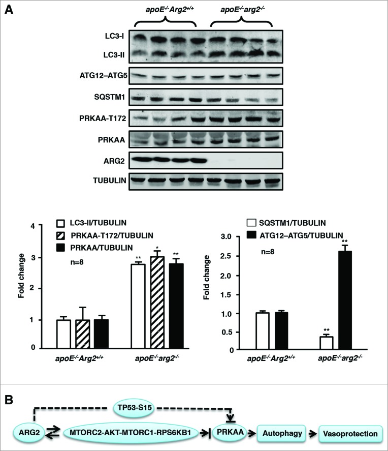
Figure 10.
Ablation of Arg2 in atherosclerosis-prone apoe−/− mice enhances PRKAA signaling and autophagy in the aortas. apoe−/−Arg2+/+ and apoe−/−arg2−/− mice were fed a high-fat diet for 10 weeks. (A) Aortas from mice of both genotypes were cleaned of perivascular tissues and subjected to immunoblotting analysis of LC3-I/-II, ATG12–ATG5, SQSTM1, PRKAA-T172 and total PRKAA, ARG2, and TUBULIN. Quantification of the signals is shown in the corresponding lower panels (n = 8 each group). *P < 0.05, **P < 0.001 vs. apoe−/−Arg2+/+. (B) Schematic summary of the major findings of this study. A feed-forward crosstalk between elevated ARG2 expression and RICTOR activates the MTORC2-AKT-MTORC1-RPS6KB1 signaling cascade, which leads to inhibition of PRKAA and subsequently inhibition of endothelial autophagy in senescent endothelial cells. ARG2 can also activate the TP53 pathway in parallel with MTOR signaling, resulting in inhibition of PRKAA and suppression of autophagy. ARG2 exerts these effects independently of its L-arginine ureahydrolase activity. The reduced PRKAA activation by ARG2 results in impairment of endothelial autophagy and acceleration of endothelial senescence and atherosclerosis.

