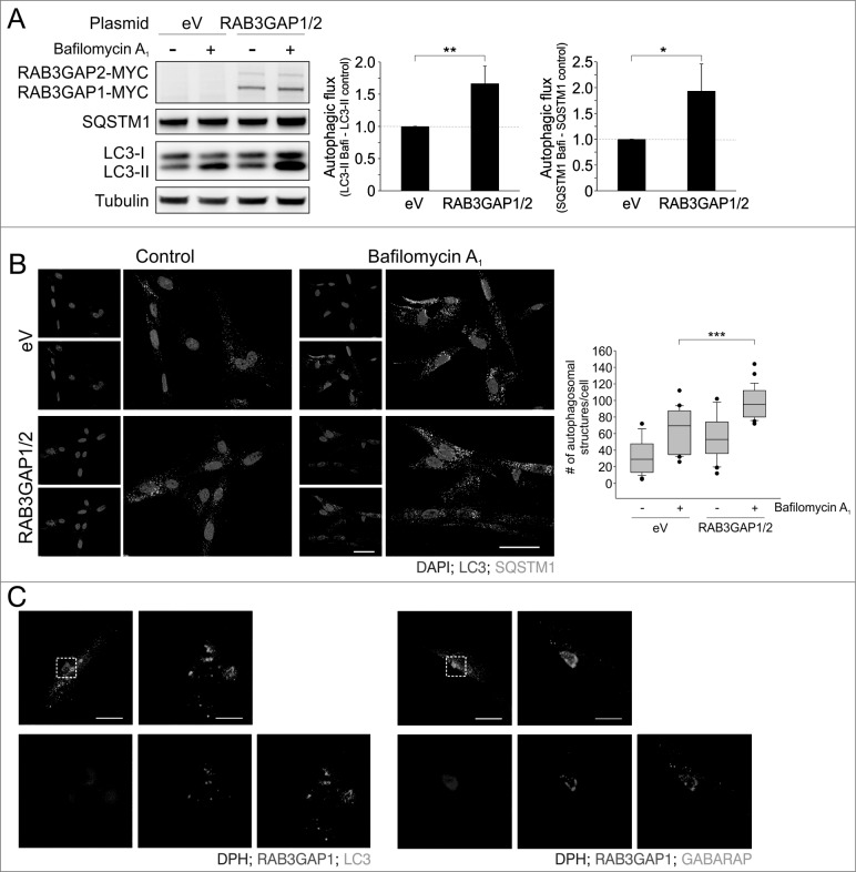
Figure 3.
Overexpression of the RAB3GAP complex enhances autophagy. (A) Immunoblot analysis of cells that were manipulated with the indicated plasmids for 48 h and treated with DMSO (−) or bafilomycin A1 (+) for 4 h. Tubulin served as control for equal loading. Statistics are depicted as mean ± SD normalized to eV; *P < 0.05, **P < 0.01, n = 4, t test. (B) Confocal images of LC3 and SQSTM1 immunostaining. Fibroblasts were manipulated with the indicated plasmids for 48 h and treated with DMSO or bafilomycin A1 for 4 h. DAPI was used to stain nuclei. Scale bar = 50 μm. Autophagosomal structures were counted in 20 to 40 cells of 3 independent experiments; ***P < 0.001, t test. (C) Confocal images of RAB3GAP1 and LC3 or GABARAP immunostainings. RAB3GAP1 and RAB3GAP2 were overexpressed for 48 h. DPH was used to stain lipid droplets. Scale bar = 20 and 5 μm.

