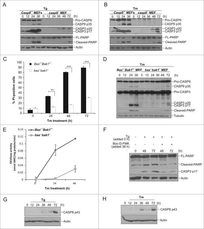
Figure 2.
ER stress-induced cell death in apoptosome-compromised cells is accompanied by caspase activation. (A and B) Casp9+/+ and casp9−/− MEFs were treated for the indicated times with (A) 0.5 μM Tg or (B) 0.5 μg/ml of Tm and lysates immunoblotted for CASP9, CASP3, PARP, and actin. (C) Bax+/+ Bak1+/+ and bax−/− bak1−/− MEFs were treated with 1 μg/mL of Tm for indicated timepoints. Cell viability was analyzed by PI uptake. (D) Bax+/+ Bak1+/+ and bax−/− bak1−/− MEFs were treated with 1 μg/ml of Tm for the indicated times and lysates were immunoblotted for CASP9, CASP3, PARP and tubulin. (E) Bax+/+ Bak1+/+ and bax−/− bak1−/− MEFs were treated with 1 μg/ml of Tm for indicated times and CASP3-like activity was determined by DEVD-AMC hydrolysis. Results are representative of at least 3 independent experiments. Error bars represent the mean ± SD. (G) casp9−/− MEFs were treated with 0.5 μM Tg with or without Boc-D-FMK for the indicated times and lysates immunoblotted for PARP and cleaved CASP3. (H and I) casp9−/− MEFs were treated with (H) 0.5 μM Tg or (I) 0.5 μg/ml of Tm for the indicated times and lysates immunoblotted for cleaved CASP8.

