Figure 1.
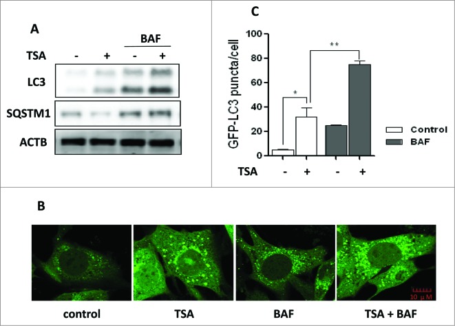
HDACIs induce autophagy. (A) HCT116 cells were treated with trichostatin A (TSA) (0.5 μM) alone or in combination with 15 nM BAF for 12 h. Cell lysates were lysed, collected, and immunoblotted using western blotting for LC3 and SQSTM1 levels. (B) MEFs with stable expression of GFP-LC3 were treated with 1 μM TSA in the presence or absence of 15 nM BAF for 12 h. The cells were examined and representative cells were photographed using a confocal microscope (Scale bar: 10 μm). (C) The number of GFP-LC3 puncta/cell was counted and presented (*P < 0.05, **P < 0.01).

