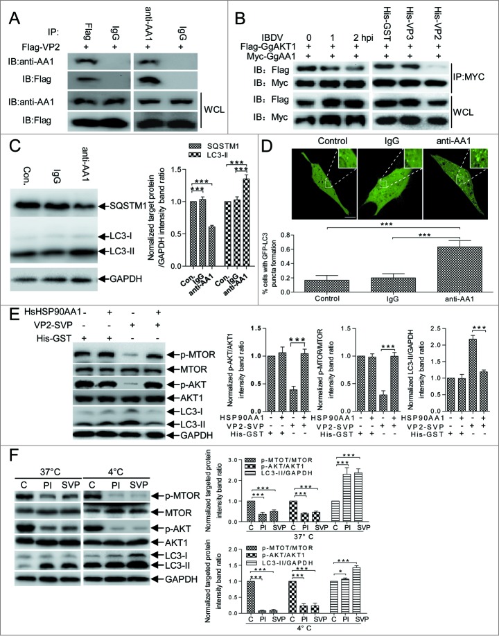
Figure 6.
HSP90AA1 binding to VP2 triggers autophagy via AKT-MTOR dephosphorylation. (A) DF-1 cells transfected with pFlag-VP2 for 24 h. Whole cell lysates (WCL) were used for CoIP and western blotting with anti-Flag or anti-HSP90AA1 antibody (anti-AA1) and irrelevant IgG (Control). (B) DF-1cells cotransfected with Myc-GgHSP90AA1 (Myc-GgAA1) and Flag-GgAKT1 for 48 h. Transfected cells were infected with IBDV for 1 or 2 h, or were incubated in DMEM containing His-VP2(100 ng/ml), His-VP3(100 ng/ml) or His-GST(100 ng/ml). Whole cell lysates of each sample were used for CoIP with anti-MYC antibody and western blotting with anti-Flag or anti-MYC antibody. (C) Western blotting performed using anti-LC3 antibody and anti-SQSTM1 mAb on lysates from DF-1 cells cultured in uncoated plates or in coated plates with anti-HSP90AA1 or irrelevant isotype control IgG for 4 h. The ratio of SQSTM1 or LC3-II to GAPDH was normalized to control conditions. (D) DF-1 cells transfected with peGFP-LC3 for 24 h and cultured in plates coated with negative control, IgG, or anti-HSP90AA1 for 4 h. Autophagic vacuoles were analyzed under confocal microscopy. The ratio of cells containing >3 ring-like GFP structures was determined. Scale bars: 10 10 μm.mu;m. Error bars: Mean ± SD of 3 independent tests. (E) DF-1 cells were incubated respectively with the His-GST, mixture of His-GST and HSP90AA1 (His-GST:HSP90AA1 = 1:2.5), mixture of SVP and HSP90AA1 (SVP:HSP90AA1 = 1:2.5) or SVP for 2 h, and analyzed by immunoblotting with anti-LC3, anti-p-MTOR, anti-MTOR, anti-p-AKT, anti-AKT1, or anti-GAPDH antibody. (F) DF-1 cells incubated with His-GST (100 ng/ml), SVP (100 ng/ml), or purified IBDV (MOI = 10 ) for 2 h at 37°C or 4°C. Cells were analyzed by immunoblotting using the antibody in (E). The ratio of p-MTOR to MTOR, p-AKT to AKT or LC3-II to GAPDH was normalized to mock infection and set at 1.0. Two-way ANOVA; ***P < 0.001 compared to control. C, His-GST; PtdIns, purified IBDV; SVP, His-VP2 subviral particle.

