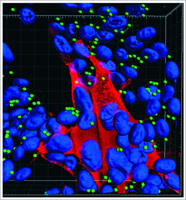
Figure 1.

Autophagosomes (green spheres) in the cytoplasm of cultured cells infected with VZV. VZV-infected monolayers were fixed with 2% paraformaldehyde, permeabilized with 0.02% Triton X-100, and then immunolabeled with a rabbit polyclonal antibody against LC3 and a mouse monoclonal antibody against the VZV major immediate-early protein IE62. Monolayers were observed with a Zeiss LSM710 Spectral confocal microscope using 63X high numerical-aperture oil immersion objective lenses. Confocal Z-stacks comprising 40 optical slices were reconstructed into 3D animations with the aid of Imaris software. The Spot function in Imaris automatically locates autophagosomes based on size and intensity thresholds; each autophagosome is represented by a sphere 500 nm in diameter. With this imaging technology, individual autophagosomes are easily detectable within infected cells. Note: VZV infection induces fusion of cells, thus there is a large cluster of nuclei in the image. Blue, Hoechst 33342 nuclear stain; red Alexa Fluor 546, VZV protein; green Alexa Fluor 488, LC3-labeled autophagosomes.
