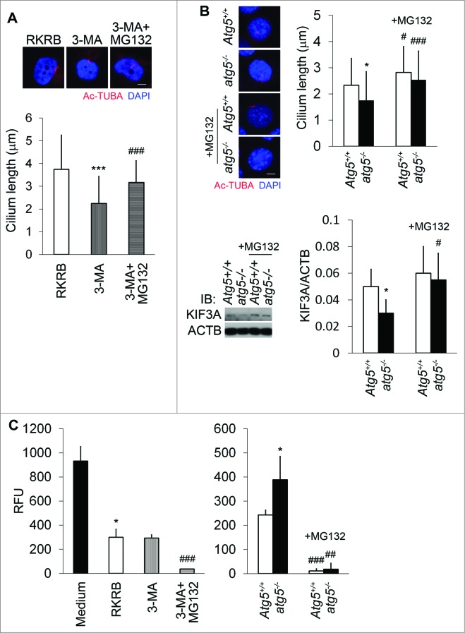
Figure 6.
Proteasome in cilia shortening in autophagy-suppressed cells. (A) Restoration of cilium length by MG132 in 3-MA-treated cells. HK2 cells were treated for 2 h with 3-MA in the absence or presence of 5 μM MG132, followed by immunofluo-rescence analysis of cilia by Ac-TUBA. Thirty six, 35, and 38 cilia were measured for the control, 3-MA-treated, 3-MA+MG132-treated cells respectively. (B) Effect of MG132 on cilium length in atg5-KO cells and induction of KIF3A by MG132. Atg5+/+ and atg5−/− cells were incubated without or with MG132 for 2 h, followed by immunofluorescence analysis of Ac-TUBA. Thirty three, 48, 46, and 50 cilia were measured for the wild-type and KO cells at the basal condition or with MG132 treatment. atg5-KO cells had shorter cilia at the basal condition. Cell lysates were collected in (B) for immunoblot analysis. Densitometry was conducted to determine the KIF3A/ACTB ratio. The results are the summary of 3 separate experiments. (C) Proteasome activity increment in autophagy-suppressed cells. Compared to Atg5+/+ cells, atg5−/− exhibited enhanced proteasome activity. MG132 significantly suppressed the proteasome activity in HK2 and Atg5+/+ and atg5−/− MEF cells. Compared to culture medium, RKRB medium reduced proteasome activity in HK2 cells. Relative fluorescent units (RFU) were used to indicate the proteasome activity in different culture conditions. Results are the summary of 4 separate experiments. P*<0.05, ***<0.001 vs RKRB (A) or Medium (C), atg5−/− vs Atg5+/+ (B); #<0.05, ##<0.01, ###<0.001 +MG132 vs -MG132 (A, B, and C). Scale bar (A and B): 5 μm.

