Figure 4.

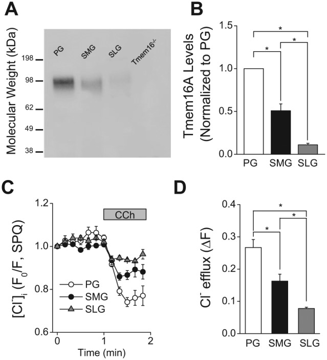
TMEM16A protein expression and muscarinic receptor-activated decrease in [Cl-]i in mouse parotid (PG), submandibular (SMG), and sublingual gland (SLG) acinar cells. (A) Western blot analysis with an anti-TMEM16A antibody was performed as described in the “Appendix Materials and Methods” section by loading 10 μg of acinar protein/lane isolated from mouse (C57BL/6) PG, SMG, and SLG. The specificity of the anti-TMEM16A antibody was confirmed by the absence of a reactive band in the lane loaded with SMG protein from a mouse lacking TMEM16A (Tmem16A-/-). (B) Summary of 4 independent experiments performed on individual protein samples obtained from 4 mice (2 females and 2 males). Band intensities (obtained by densitometric analysis with ImageJ software) were normalized by the intensity obtained from PG in each separate blot, which displayed the highest expression level among major salivary glands. (C) Changes in [Cl-]i in response to 0.3 µM CCh were measured in SPQ-loaded cells from mouse (C57BL/6) PG, SMG, and SLG. (D) Summary of the magnitude of the [Cl-]i decrease in PG, SMG, and SLG acinar cells from mouse (C57BL/6). The [Cl-]i decrease was significantly greater in the PG, followed by the SMG and then the SLG. One-way ANOVA followed by Bonferroni’s post hoc test was applied for the detection of statistically significant differences. *P < 0.05. n = 4 (males = 2, females = 2).
