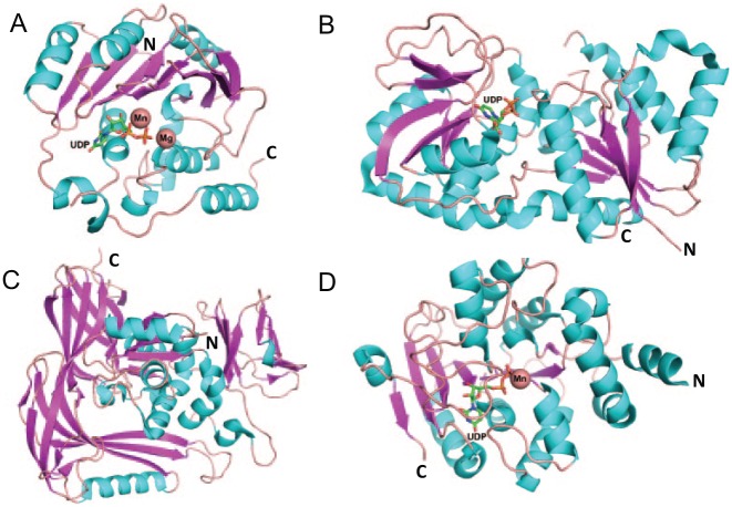
Figure 1.
Structural folds of glycosyltransferases (GTs). Structural ribbon diagrams are colored: purple for β-sheet, cyan for helix, and pink for loops. GT-A fold structure, SpsA (PDB entry, 1QGQ) (A); GT-B fold structure, Gtf3 (PDB entry, 3QKW) (B); GT-C fold structure, STT3 (PDB entry, 2ZAG) (C); GT-D fold structure, DUF1792 (PDB entry 4PHR) (D). Uridine diphosphate is found in 3 glycosyltransferase folds, GT-A, B, and D. Mn and Mg are shown in the sphere.

