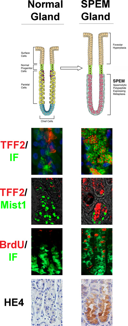
Figure 1.
Comparison of normal fundic gastric glands and metaplastic SPEM glands in mice. The diagram at the top depicts alterations in gland lineages between normal and SPEM glands with emergence of SPEM and foveolar hyperplasia following loss of parietal cells. Panels below show immunostaining patterns for normal and SPEM-containing glands. TFF2 (red) expressing mucous neck cells redifferentiate into intrinsic factor (green) expressing chief cell at the base of normal glands. However, TFF2 expression is expanded to the base of SPEM glands and appears within cells that show dual staining for intrinsic factor. Mist1 (green) is a differentiated chief cell marker in normal gastric glands. However, Mist1 is also expressed in some TFF2-expressing SPEM cells, suggesting transdifferentiation of chief cells. Although proliferation as detected by BrdU (red) is normally only in the progenitor zone of the upper normal gland, TFF2-expressing SPEM cells show clear proliferating cells also expressing intrinsic factor (IF). Previous studies have led to the identification of promising biomarkers of SPEM such as HE4. HE4 is not detected in normal chief cells or any normal fundic cells, but HE4 staining is strongly observed in SPEM.

