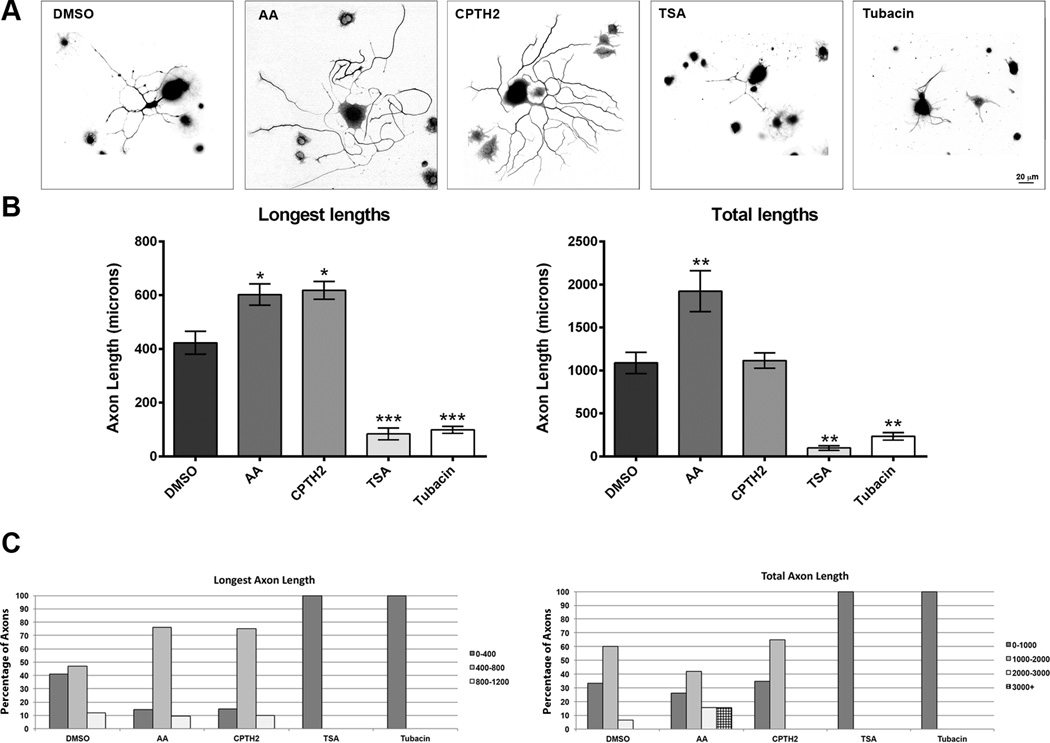
Fig 2. The HATis AA and CPTH2 significantly increase axon growth while HDACis TSA and Tubacin significantly decrease axon growth.
Dissociated DRG neurons were grown in the presence of 10 µM Anacardic acid, 50 nM CPTH2, 10 µM Tubacin, or 300 nM TSA for 48 hrs before fixing and labeled with with β-III-tubulin. A: Representative images of neurons labeled for β-III-tubulin antibody showing all the axons emerging from the cell body. B: In neurons treated with AA, the mean lengths of the longest axon and the total axon lengths were significantly longer than control neurons treated with DMSO. In neurons treated with CPTH2, only the mean length of the longest axon was significantly longer than neurons treated with DMSO. The mean total axon length was not different from control DMSO. In neurons treated with TSA and Tubacin, the mean axon lengths and total axon lengths were shorter than in neurons treated with DMSO. C: In neurons treated with AA and CPTH2, a greater proportion of neurons grew their longest axons between 400 and 800 µm than in DMSO control. In neurons treated with AA, a greater proportion of neurons grew total axon lengths that reached over 2000 µm compared with control. In neurons treated with CPTH2, there was little difference in the total axon lengths with neurons treated with DMSO. In neurons treated with TSA and Tubacin, no axons were able to grow beyond 400 µm by the end of the experiment. * p<0.05, **p<0.01, ***p,0.001.

