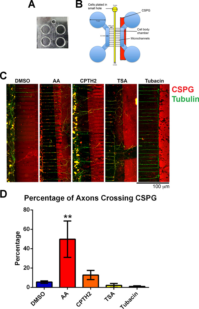
Fig 4. Neurons treated with HATis grow over inhibitory CSPG borders in microfluidic chambers while neurons treated with HDACis turn at the border.
Dissociated DRG neurons were plated into microfluidic chambers and allowed to grow axons towards a CSPG stripe. Cultures were fixed after 5 days growth and labeled with tubulin (green) and with CS-56 antibody (red) A: Microfluidic chambers were caste by pouring silicone elastomer into a pre-fabricated microfluidic template and holes were punched to mark the chambers (see Materials and Methods). B: Microfluidic chambers were placed over a CSPG stripe before neurons were plated into a small hole and allowed to occupy the Cell body chamber. Neurons were grown for 5 days until axons reached the CSPG stripe. C: In neurons treated with AA, a significant proportion of axons were able to cross the CSPG border compared with neurons treated with DMSO. In neurons treated with CPTH2, more axons were able to cross the CSPG border compared with neurons treated with DMSO, but this proportion was less compared with DMSO. In neurons treated with TSA and Tubacin, very few axons were able to cross the CSPG border. **p<0.01.

