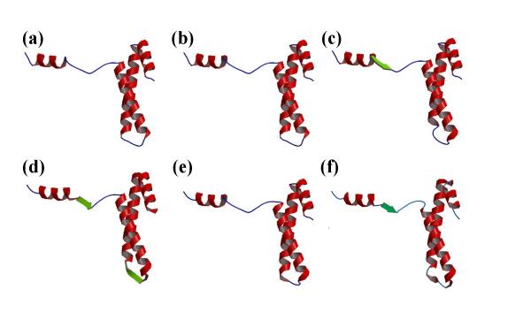
Figure 3.

Representation of the secondary structure assignments. Example of the ribosomal protein S15 from Bacillus Stearothermophilus (PDB code 1A32) with (a) DSSP, (b) STRIDE, (c) PSEA, (d) DEFINE, (e) PCURVE and (f) Protein Blocks. In the last case to simplify the representation, helices are associated to PB m and strands to PB d. The visualization is done with RASTER 3D [42,43] and MOLSCRIPT [44]. The α-helices are in red, the β-strands in green and the coils in blue.
