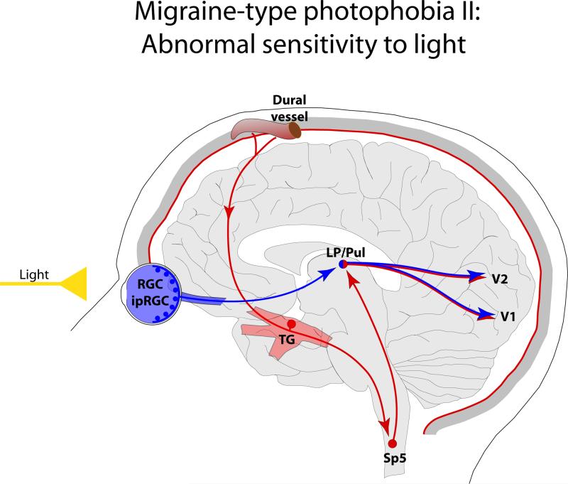
Figure 2.
Proposed mechanism for enhanced sensitivity to light during migraine through the convergence of nociceptive signals from the meninges on thalamic neurons that project to the visual cortices. Red depicts the trigeminovascular pathway. Blue depicts visual pathway from the retina to the visual cortex. Abbreviations: V1, primary visual cortex; V2 secondary visual cortex. For other abbreviations see Fig. 1.

