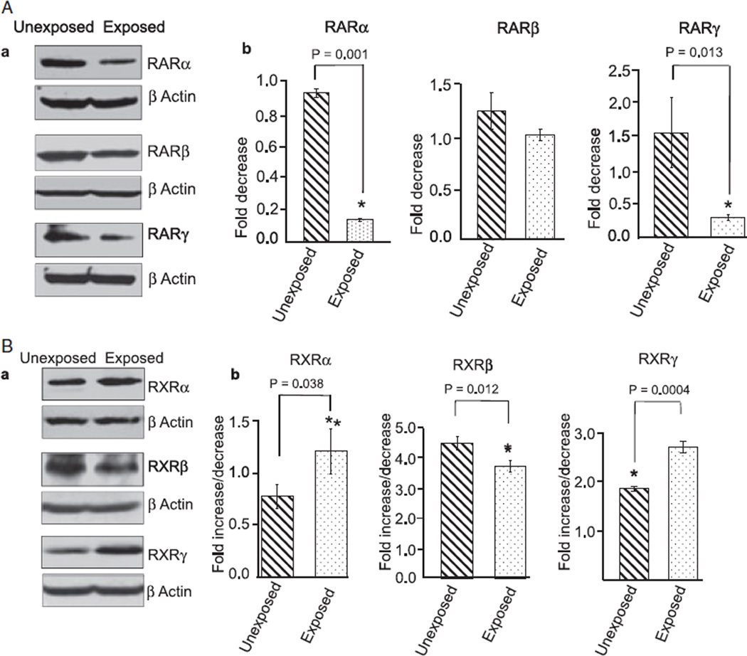
Fig. 2.
Ethanol changes the expression of RA receptors in the cerebellum, as shown by Western blot analysis. Cerebella of unexposed and ethanol-exposed pups were homogenized in RIPA buffer for protein extraction. Equal amount of extracted proteins from the unexposed and ethanol-exposed samples was separated by SDS–PAGE and transferred to a nitrocellulose membrane for Western blotting using anti-RA receptor antibodies. (A) Western blot analysis of cerebellar proteins using anti-RARα, β, and γ antibodies. (B). Western blot analysis of cerebellar proteins using anti-RXRα, β, and γ antibodies. Membranes were re-probed with an anti-actin antibody to examine loading differences. The expression of RARs and RXRs was quantified by scanning densitometry of their respective bands. The ratio of the scanned density of the band belonging to the individual receptor with that of the actin is represented as fold increase or decrease. Results are mean ± SD of three separate experiments.

