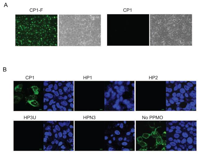
Fig. 3. PPMO enter S10-3 liver cells and inhibit HEV replication.
A. PPMO uptake assay in S10-3 cells. Fluorescein-conjugated CP1 (CP1-F) or CP1 without fluorescein conjugation were added to S10-3 cells and incubated for 4 h before fluorescence microscopy. Green fluorescence indicates uptake of PPMO. Bright field illuminations are shown next to the fluorescence images. B. Immunofluorescence assay of S10-3 cells infected with HEV. Cells were transfected with Sar55 RNA transcribed from pSK-E2, treated with indicated PPMO (16 μM) 5 hours later, and fixed for IFA at 7 days post-transfection. In each panel, the left image shows IFA using HEV-specific antibody, and the right image shows the same field with cell nuclei stained by DAPI. A green scale bar in the lower right corner represents 10 μm.

