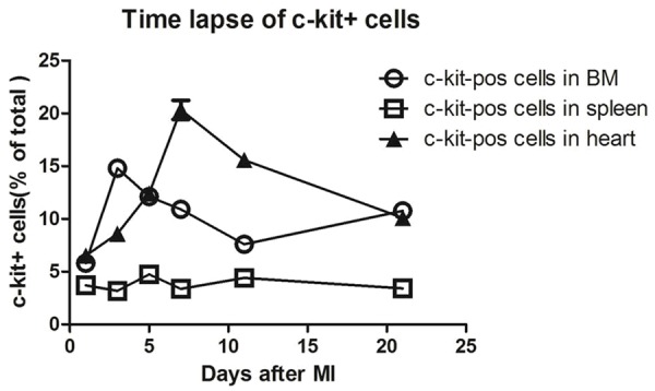
Figure 1.

The time lapse of c-kit-positive cells in mouse BM, spleen and heart after MI. Quantification of the number of c-kit+ cells over a time course after MI in wild-type mice showed that MI leads to an increase in c-kit+ first in bone marrow and then specifically within the infarcted myocardium.
