
Figure 2.
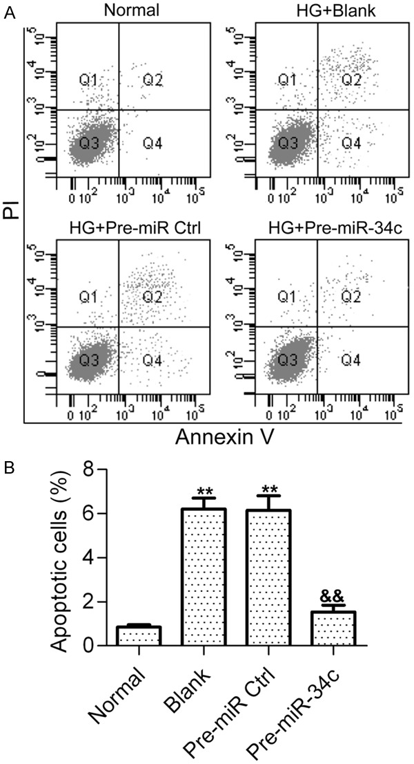
Effect of miR-34c on HG-induced podocyte apoptosis. A. Cell apoptosis was detected by flow cytometry after HG stimulation for 48 h. Podocytes treated with normal glucose (5 nM) was used as the control. Podocytes stimulated with HG were treated with PBS (Blank), control pre-miRNAs (pre-miR Ctrl) or pre-miR-34c. Podocytes were harvested and stained with Annexin V and PI before apoptotic cells were examined. B. Representative histograms show the fractions of apoptotic cells. Statistical analysis was calculated by one-way ANOVA. N = 3, **P < 0.01 vs. Normal, &&P < 0.01 vs. Blank or Pre-miR Ctrl.