
Figure 1.
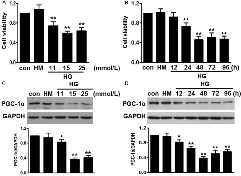
High glucose-induced cell injury was associated with decreased PGC-1α expression. A. HUVECs were incubated with different concentrations of high glucose (HG) for 48 h. Cell viability was analyzed by cell counting kit-8 (CCK-8). B. The cells were treated with high glucose (15 mmol/L) for the indicated times. CCK-8 analysis was performed. C. Western blot showed that glucose inhibited PGC-1α expression dose-dependently. D. After treatment mentioned in B, PGC-1α protein level was determined by western blot. All data were expressed as mean ± SEM. *P < 0.05, **P < 0.01 vs. control, n = 6.