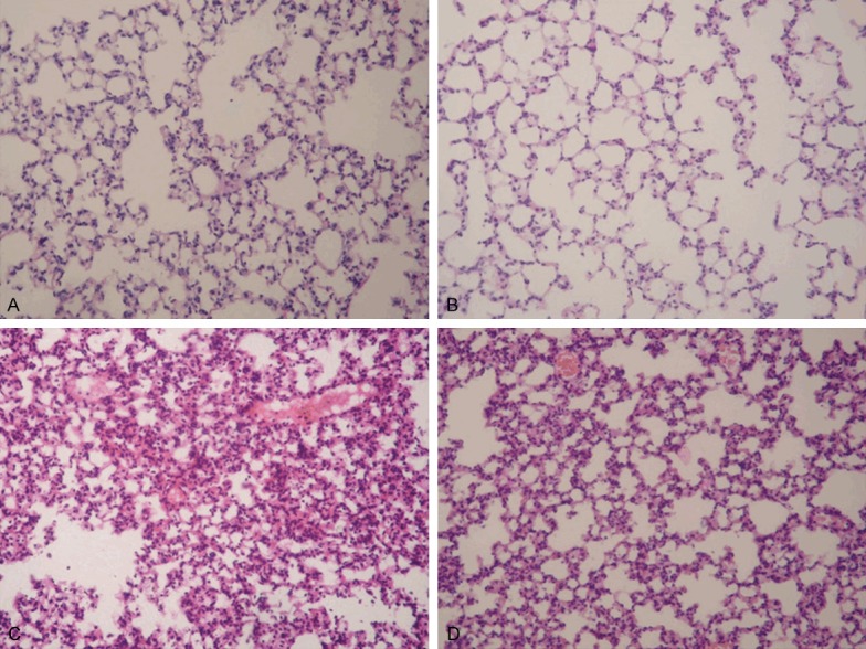
Figure 1.

Effect of crocin on the pulmonary histopathological changes of mice with ALI. Lung sections stained with hematoxylin-eosin from 12 h after LPS administration revealed pulmonary histopathological changes (original magnification ×200). A. Control group: normal structure. B. Crocin group: same as control group. C. LPS group: alveolar wall thickness, hemorrhage, alveolus collapse and obvious inflammatory cells infiltration. D. LPS+crocin group: minor histopathological changes compared with LPS group.
