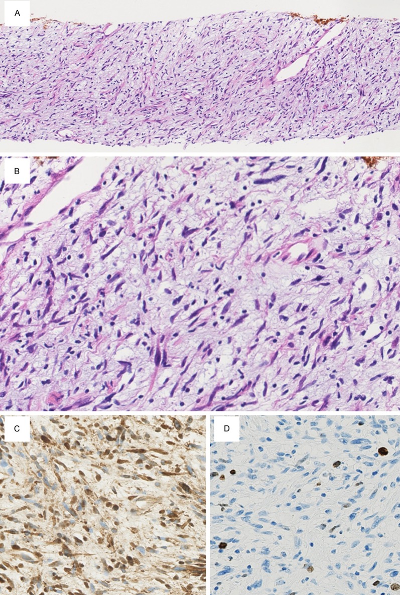
Figure 2.

Microscopic findings of the biopsy specimen. A. Cellularity of the specimen is higher than that of a neurofibroma and the Antoni A areas of a schwannoma (×40). B. Some spindle cells show nuclear enlargement and hyperchromasia (×400). Mitotic figures are not observed. C. Numerous spindle cells are S-100 positive on immunohistochemistry (×400). D. The Ki-67 labeling index is approximately 6.4% (×400).
