
Figure 3.
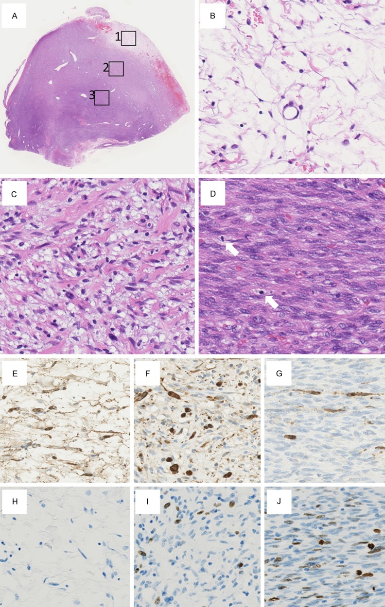
Microscopic findings of the surgically resected specimen. (A) A surgically resected specimen shows 3 components correlating with their cellularity, which show gradual transition from a low cellular component (boxed area 1) to a moderate cellular component (boxed area 2) to a high cellular component (boxed area 3) (×12.5). (B) High-power view of the boxed area 1 in (A). (A) component comprising spindle cells with low cellularity, no apparent nuclear atypia, and no mitotic figures was found at the periphery of the tumor (×400). (B) High-power view of the boxed area 2 in (A). (A) component consisting of spindle cells with moderate cellularity and mild nuclear pleomorphism as well as nuclear hyperchromasia is observed (×400). (D) High-power view of the boxed area 3 in (A). (A) component displaying fascicular growth pattern of spindle cells with high cellularity is shown, constituent cells of which show moderately enlarged nuclei and coarse chromatin. Two mitotic figures (arrows) are observed in this field (×400). (E) On immunohistochemistry, the neurofibroma is diffusely positive for the S-100 protein (×400). (F) On immunohistochemistry, the low-grade malignant peripheral nerve sheath tumor is sparsely positive for the S-100 protein (×400). (G) On immunohistochemistry, the high-grade malignant peripheral nerve sheath tumor is rarely positive for the S-100 protein (×400). (H) The Ki-67 labeling index is less than 1% in the neurofibroma (×400). (I) The Ki-67 labeling index is 7.8% in the low-grade malignant peripheral nerve sheath tumor (×400). (J) The Ki-67 labeling index is 21.6% in the high-grade malignant peripheral nerve sheath tumor (×400).