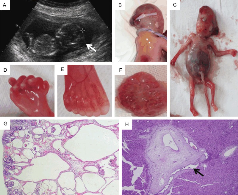
Figure 1.

Clinical features of two affected fetuses in Chinese MKS3 family. (A) Ultrasound scans of III-3 at 14 gw. Occipital encephalocele was observed and confirmed by autopsy for III-3 (B). Parietal absence, distended abdomen due to enlarged kidney, clearly visible cysts and polydactyly of the feet were observed in III-4 fetus (C-F), which is rarely seen in MKS3 fetuses. The kidney of III-4 (F) showed diffuse multicystic renal dysplasia. Cysts are found in the deep cortex and medulla, smaller at the periphery than in the center; notice the remnant glomerulus in renal cortex (G). The liver biopsy showed hepatic ductal plate malformations. Enlarged intrahepatic bile duct is indicated by the arrow (H).
