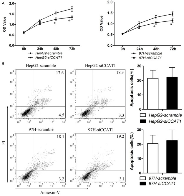
Figure 2.
Effects of CCAT1 on proliferation and apoptosis in HCC cell lines. A. CCK-8 cell proliferation assays show that CCAT1 knock-down significantly weakened proliferation in HepG2 and MHCC-97H cells. Data are represented as mean ± SEM. *Indicates P < 0.05, independent experiment was performed three times. B. Representative flow cytometric plots of cell apoptosis. Cells were stained with both Annexin V and PI before analysis by flow cytometry. Numbers represent the frequency in each quadrant. Data are represented as mean ± SEM. *Indicates P < 0.05, independent experiment was performed three times.

