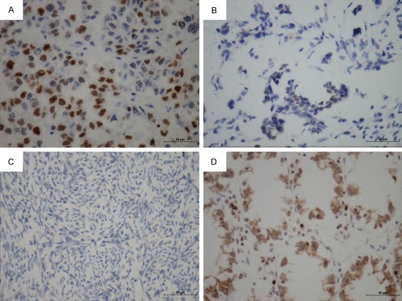
Figure 1.

Expression of pH2AX in EOC and normal ovarian tissue. A. High expression of pH2AX in clear cell carcinoma (×400). B. Low expression of pH2AX in serous carcinoma (×400). C. Negative expression of pH2AX in normal ovarian tissue (×400). D. Cytoplasm staining was also evident in some cancer cells (mucous carcinoma, x400).
